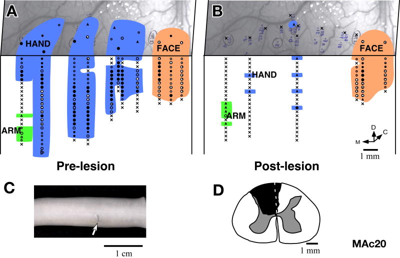
Fig. 16.
Somatotopy in area 3b of monkey MAc20 before and immediately after lesion of the dorsal columns. (A) Locations of neurons with receptive fields on the face, hand and arm encountered in area 3b and adjacent cortex before, and (B) immediately after lesion of the contralateral dorsal columns of the spinal cord. Post-lesion only a few neurons responded weakly to hard taps on the hand or arm. The photograph underlay in ‘A’ and ‘B’ shows the correspondence of the pre- and post-lesion penetration sites with respect to the surface vasculature of the brain. (C) Photograph showing lesion (arrow) on the dorsal surface of the spinal cord. Rostral is to the left. (D) The lesion reconstructed from the transverse sections of the spinal cord.

