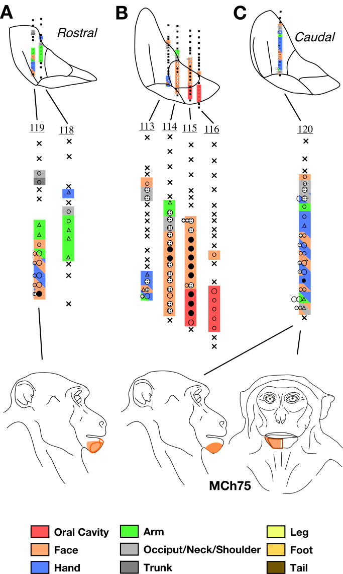
Fig. 17.
Somatotopy and receptive fields encountered in seven electrode penetrations through the ventroposterior (VP) nucleus of monkey MCh75 shown on a series of three rostral to caudal coronal sections (A, B and C). The figurines at the bottom show abnormally located receptive fields on the face for neurons in the lateral portion of VP. Normally located receptive fields i.e. those on the face in VPM, and other body parts in VPL are not shown. For illustration purposes, electrode penetrations within a rostrocaudal extent of 500μm are projected on a single intermediate section. The penetrations are numbered. The color key shown at the bottom also applies to the maps shown in figures 18-23 and is same as used for illustrating maps of area 3b. The regions shown in double or triple colors by use of colored slanting lines indicate mapping sites where neurons had multiple receptive fields on body parts indicated by the colors of the lines. For other conventions see Fig. 4A.

