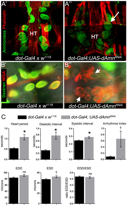
Figure 4. Loss of Amnionless function in nephrocytes leads to cardiomyopathy.
(A) Amnionless was silenced in nephrocytes using dot-Gal4. As a negative control, the parent driver line was outcrossed once to the w1118 line (dot-Gal4 × w1118) and offspring analysed. Micrographs show adult hearts stained with anti-Amnionless antibodies (green) and phalloidin (red). Amnionless protein was localised to nephrocytes in controls (A') whereas silencing led to reduction in its detection but not the loss of pericardial nephrocytes (A”; arrow indicates a pericardial nephrocyte); HT = Heart tube. (B) Semi-intact heart preparations were incubated with fluorescently-tagged 10kDa dextran (green) and wheat germ agglutinin (red) for 30 minutes, washed and imaged. Arrows indicate nephrocytes. Dextran accumulated in controls (B') but not in Amnionless-silenced nephrocytes (B”). WGA = wheat germ agglutinin. (C) Quantification of heart function in flies with dAmnionless silenced nephrocytes. Hearts were analysed by high frame rate videomicroscopy of semi-intact adult heart preparations. EDD = end diastolic interval; ESD = end systolic diameter; EDD/ESD = fractional shortening of the heart contraction; ns = not statistically different from control genotype (dot-Gal4 × w1118); *P<0.01, †*<0.05; n = 18-20 per genotype.

