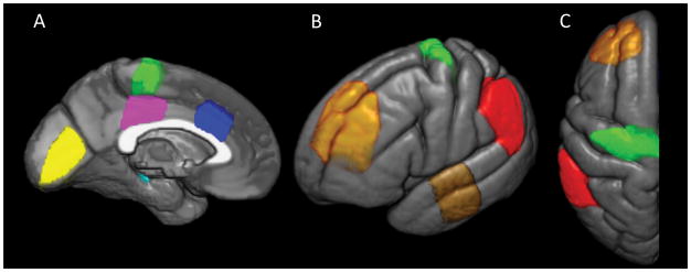
Figure 2. Atlas ROIs overlaid on the left hemisphere of a common template.
Medial (A), lateral (B), and axial (C) views of the atlas ROIs. Yellow = primary visual cortex, Cyan = posterior hippocampus, Magenta = posterior cingulate gyrus, Green = primary motor cortex, Blue = anterior cingulate gyrus, Orange = midfrontal gyrus, Brown = superior temporal gyrus, Red = inferior parietal lobule.

