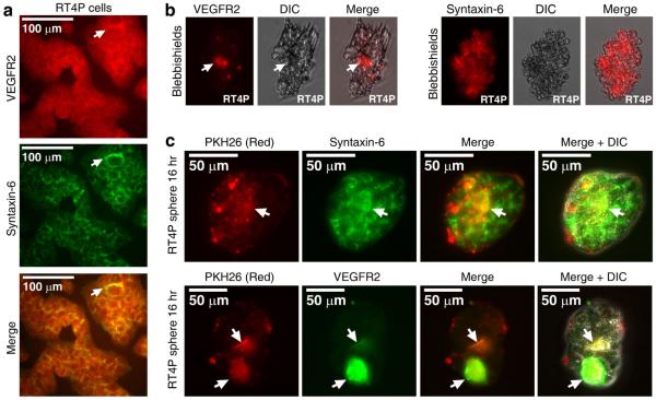
Fig. 5. VEGFR2 undergoes endocytosis in blebbishields, and internalized VEGFR2 colocalizes with PKH26-labelled lipid membranes in transformed spheres.
a-b, Double immunofluorescence analysis of VEGFR2 and syntaxin-6 (trans-Golgi-network marker) showed their co-localization at the perinuclear trans-Golgi-network (arrows) in non-apoptotic cells (a). Immunofluorescence analysis of VEGFR2 and syntaxin-6 and in freshly fixed blebbishields; internalized VEGFR2 is marked by arrows (b). c, Lipo-immunofluorescence analysis of VEGFR2 and syntaxin-6 co-localization with PKH26-labelled blebbishield surface membrane lipids (internalized in spheres; co-localization marked by arrows). DIC, differential interference contrast.

