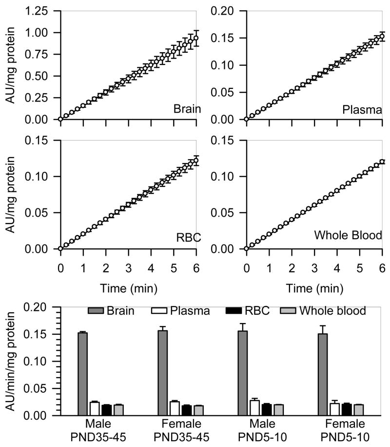
Figure 3. Rate of AChE hydrolysis of acetylthiocholine in different tissues from male and female guinea pigs at PND5-10 and PND35-45.
As described in Methods, AChE activity was measured by means of a colorimetric assay (Ellman et al., 1961) in brain extracts, RBC, plasma and whole blood from neonatal and prepubertal guinea pigs of both sexes. A kinetic profile of the enzyme activity (expressed as the changes in absorbance units (AU)/mg protein) was studied spectrophotometrically at 15-s intervals. Scatter graphs show the time changes in absorbance units/mg protein in brain extracts, plasma, RBC, and whole blood pooled from six prepubertal male guinea pigs. Each experiment was carried out in triplicate. Data points and error bars represent the mean and SEM of the actual measurements at any given time. The bar graph shows the rate of acetylthiocholine hydrolysis, estimated as the slope of the plots of AU/mg protein vs. time, in brain extracts, plasma, RBC, and whole blood from neonatal and prepubertal guinea pigs of both sexes. Bar graphs and error bars are mean and SEM of results obtained from triplicate experiments. In all animal groups, AChE activity was significantly higher in the brain extracts than in any blood compartment. However, the total enzyme activity was not sex or age dependent.

