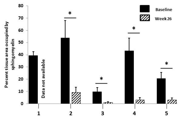
Figure 1. Metamorph quantification of sphingomyelin in liver biopsies pre- and post-olipudase alfa treatment.

This staining and quantification method identifies and measures only abnormal sphingomyelin accumulation which is part of the disease process. The normal value in a non-ASMD liver is 0. The “n” for number of blocks analyzed by Metamorph at baseline and week 26, respectively, for each patient are as follows: Patient 1, n=2, n=0; patient 2, n=4, n=8; patient 3, n=8, n=9; patient 4, n=5, n=6; patient 5, n=7, n=7. *p < 0.0001.
