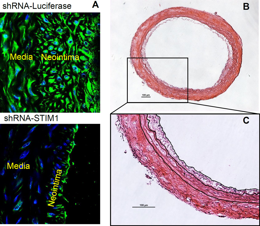
Figure 2.
Cross sections of rat carotid arteries with treatment of shRNA-STIM1 or shRNA control, two weeks post injury. A. Immunofluorescent staining of STIM1 and DAPI staining of nuclei on cross sections from both groups. The attenuated expression of STIM1 and neointima formation exhibit in the shRNA-STIM1 treated artery section. B. H&E staining of the shRNA-STIM1 artery section. C. Digital tracing of the neointima border, IEL and EEL, for the purpose of measuring areas of lumen, neointima and media. Scale bar are 100 µm.

