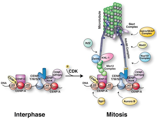
Figure 3. Attachment.
Diagram showing kinetochore structure and organization during interphase and mitosis. At mitotic entry, CDK/Cyclin B phosphorylation promotes outer kinetochore assembly upon a platform of constitutive kinetochore proteins. For information on the additional kinetochore proteins shown in the figure, see (Cheeseman and Desai 2008).

