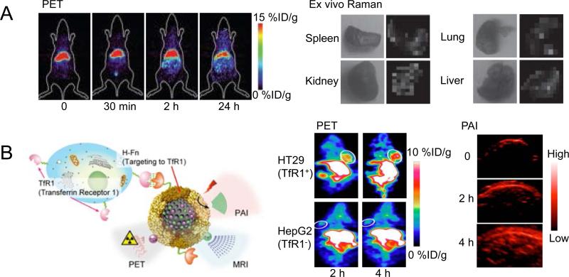
Figure 4.
(A) PET and ex vivo Raman imaging to evaluate the organ distribution of 64Cu-labeled gold nanoparticles. Consistent organ uptake was obtained by PET and Raman signals. Adapted with permission (93). (B) 64Cu-labeled melanin nanoparticles were used for tumor detection via PET and PAI. The schematic structure of MNPs is provided along with examples of both PET and PAI images of tumor-bearing mice. Adapted with permission (96).

