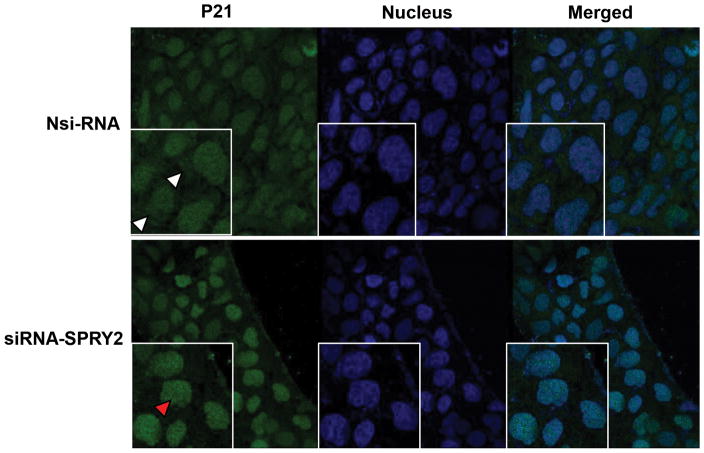
Fig. 6. Increased nuclear localization of p21 in SPRY2 downregulated cells.
Representative confocal images showing expression of p21 in Nsi RNA or SPRY2 siRNA (50 nM, 72hr) treated Caco-2 cells. Cells were fixed with cold methanol, permeabilized, and blocked with normal goat serum. Sequentially, the cells were incubated with rabbit anti-p21 and Alexa Fluor 488-conjugated goat anti-rabbit immunoglobulin. The cells were then stained with DAPI to label the nuclei. Images were obtained using a Leica TCS SP8 confocal laser scanning microscope. Cytoplasmic (white arrow head) and nuclear (red arrow head) localization of p21 is shown. All images were collected using identical microscope settings and magnification. The panels are representative of at least three independent experiments in which similar patterns were obtained for the indicated cells.

