
Fig. 1. HFD results in decreased lymphatic vascular density in obesity-prone mice.
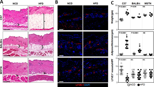
A. Representative H&E stained sections of back skin from NCD- or HFD-fed C57BL/6J, BALB/cJ, and MSTNln mice (Scale bar=200μm; bracket surrounds subcutaneous adipose tissues).
B. Representative immunofluorescent localization of LYVE-1+ vessels in upper limb tissues of NCD- or HFD-fed mice in all groups (Scale bar=100μm).
C. Upper panel: Body weights of mice on NCD (open circles) and HFD (filled circles) in all groups (n=5/group). C57BL/6J HFD vs. NCD (*p<0.0001). BALB/cJ HFD vs. NCD (*p<0.05), MSTNln mice had no significant difference. Middle panel: Quantification of subcutaneous soft tissue thickness in NCD- and HFD-fed mice in all groups (n=5–10/group). C57BL/6J HFD vs. NCD (*p<0.0001). MSTNln and BALB/cJ mice show no significant difference between NCD and HFD groups. Lower panel: Quantification of upper limb LYVE-1+ lymphatic vessel density per high powered field (HPF) and quadrant in NCD- and HFD-fed mice in all groups (n=5animals*4HPF/group). C57BL/6J HFD vs. NCD (*p<0.0001). MSTNln and BALB/cJ mice show no significant difference between NCD and HFD groups.