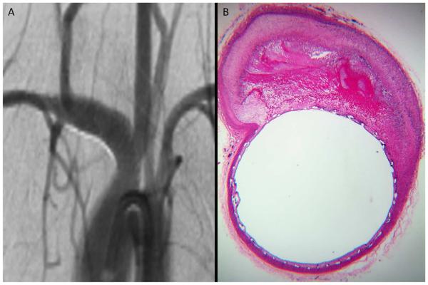
Figure 1. Histology and DSA illustrative correlation of good wall apposition associated with complete aneurysm occlusion.
Follow-up DSA objectives complete occlusion of the aneurysm sac (A). Photomicrograph (Hematoxylin-eosin stainiing, original magnification x100) at the level of the aneurysm neck shows perfect PED wall apposition with complete aneurysm pouch occlusion filled with conjunctive tissue (B).

