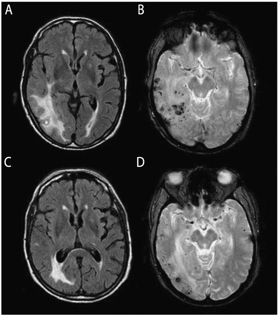
FIGURE 7-4.
MRI findings of cerebral amyloid angiopathy–related inflammation. An elderly man presented with relatively acute onset left hemiparesis, left homonymous hemianopia, dysarthria, spatial and temporal disorientation, sensory aphasia, and psychomotor slowness. Cerebral amyloid angiopathy–related inflammation was suspected and the finding of APOE genotype ε4/ε4 supported the diagnosis. Anti–amyloid-β (Aβ) autoantibody concentration in CSF was elevated at 55.9 ng/mL. Physical therapy and corticosteroid therapy with dexamethasone 24 mg/d were started and the patient showed clinical improvement. Initial axial fluid-attenuated inversion recovery (FLAIR) MRI shows bilateral hyperintense lesions (A) and gradient recalled echo (GRE) image shows cortical and subcortical microhemorrhages (B). After 1 month of steroid therapy, FLAIR MRI (C) and GRE (D) sequence show reduction of both cerebral edema and microhemorrhages.
Modified with permission from Crosta F, et al, Case Rep Neurol Med.31 www.hindawi.com/journals/crinm/2015/483020/. © 2015 Francesca Crosta et al.

