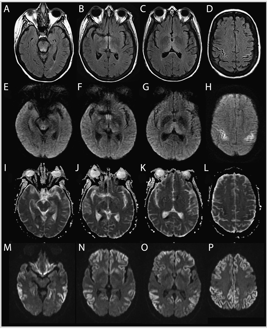
FIGURE 7-6.
MRI of the patient in Case 7-1. Wernicke encephalopathy (compared to a sporadic Jakob-Creutzfeldt disease case). Fluid-attenuated inversion recovery (FLAIR) (A–D), diffusion-weighted imaging (DWI) (E–H), and apparent diffusion coefficient (ADC) map (I–L) sequences showing FLAIR and DWI hyperintense signal changes involving the periaqueductal gray and midbrain tectum, medial thalami, and perirolandic cortex in the patient with Wernicke encephalopathy. There is relative sparing of the mammillary bodies across all sequences (B,F,J). The ADC sequences (I–L) primarily show subtle hypointensity in the perirolandic cortex (L), corresponding to hyperintensities on FLAIR (D) and DWI (H). This pattern preferentially involving the perirolandic cortex is the opposite of what we typically see in sporadic Jakob-Creutzfeldt disease (M–P; DWI sequences), in which there is generally sparing of the perirolandic region, particularly the primary motor cortex.

