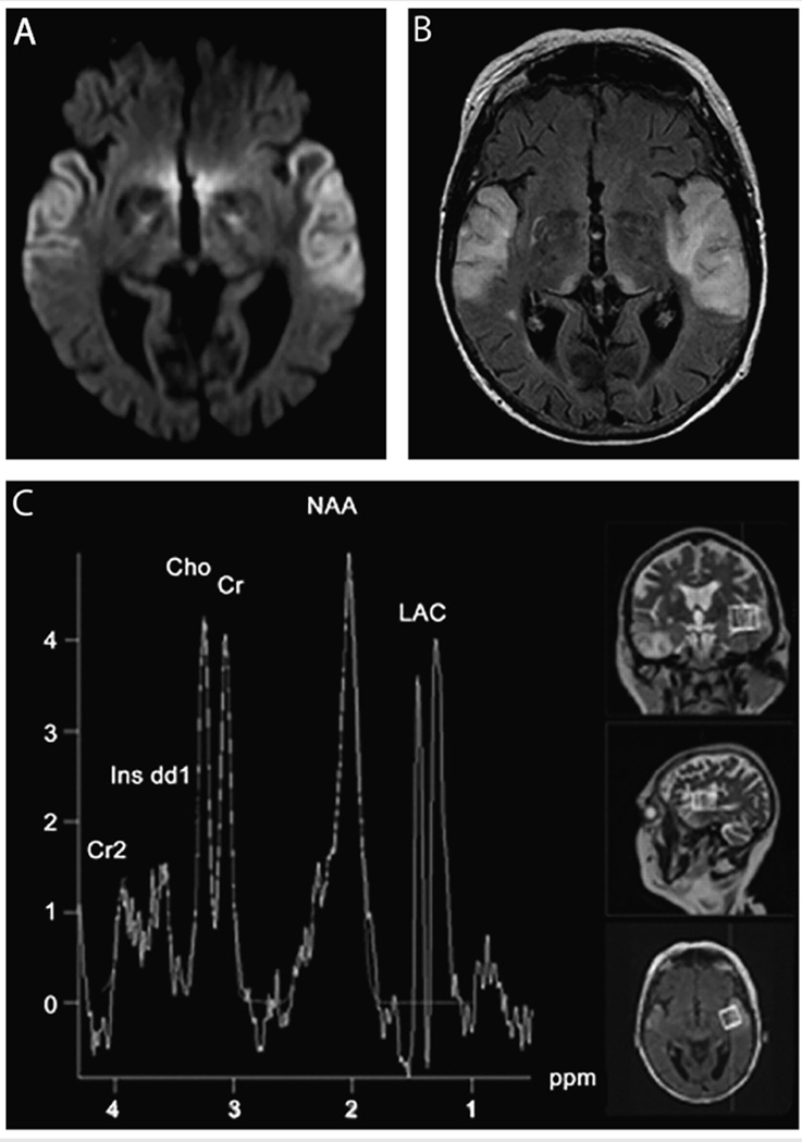
FIGURE 7-9.
MRI and spectroscopy in a case of mitochondrial myopathy, encephalopathy, lactic acidosis, and strokelike episodes syndrome (MELAS). A 59-year-old woman developed confusion, progressive aphasia, mutism, and fluctuations of alertness over 2 weeks. Diffusion-weighted imaging (DWI) MRI revealed abnormalities overlapping with Jakob-Creutzfeldt disease (A), although the fluid-attenuated inversion recovery (FLAIR) MRI (B) with white and gray matter hyperintensity was not consistent with Jakob-Creutzfeldt disease. CSF showed normal cell counts, negative polymerase chain reaction (PCR) for herpes simplex virus, elevated lactate (4.6 mmol/L), and increased levels of 14-3-3 and tau protein (1300 pg/L), both concerning for Jakob-Creutzfeldt disease. There were no periodic sharp-wave complexes on EEG recordings. Magnetic resonance spectroscopy revealed a lactate signal indicative of mitochondriopathy and genetic analysis confirmed the MELAS A3243G mutation. The DWI (A) displays bitemporal neocortical hyperintense signals. The FLAIR (B) 2 days after the initial MRI scan reveals newly emerging symmetric lesions in the pulvinar thalami. Magnetic resonance spectroscopy (C) displays a strong lactate signal.
Cho = choline; Cr = creatine; Cr2 = phosphocreatine; Ins dd1 = myoinositol; LAC = lactate; NAA = N-acetylaspartate; ppm = parts per million.
Reprinted with permission from Weiss D, et al, Neurology.59 www.neurology.org/content/77/9/914.full. © 2011 American Academy of Neurology.

