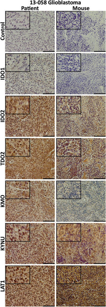
Figure 3.
Immunostaining for kynurenine pathway’s (KP) elements in patient and xenograft tumor tissues. Immunohistochemical staining for secondary antibody only control; the rate-limiting enzymes indoleamine 2,3-dioxygenase (IDO) 1, IDO2, and tryptophan 2,3-dioxygenase (TDO2); downstream enzymes kynureninase (KYNU) and kynurenine 3-monooxygenase (KMO); and l-type amino acid transporter 1 (LAT1), the membrane transporter that mediates the uptake of TRP and AMT. Left column: 13-058 patient tumor tissue and right column: mouse xenograft tumor tissue. Staining patterns are parallel between the 2 tumors with stronger signals found from IDO2 and TDO2 and the weakest signals found from IDO1 and KMO. The length of the scale bar is 100 µm. Insets: 4× greater magnification than the larger image.

