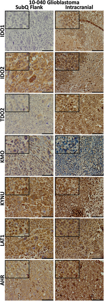
Figure 6.
Immunohistochemical staining of the 10-040 patient-derived xenograft (PDX) models. Tissues were collected from the mouse subcutaneous (subQ) flank tumor (left) and the intracranial (right) mouse models. Tissues were stained for the kynurenine pathway (KP) enzymes, l-type amino acid transporter 1 (LAT1), and the transcription factor aryl hydrocarbon receptor (AHR). For all except kynurenine 3-monooxygenase (KMO), staining was equivalent or greater in the intracranial tumor. The length of the scale bar is 100 µm. Insets: 4× greater magnification than the larger image.

