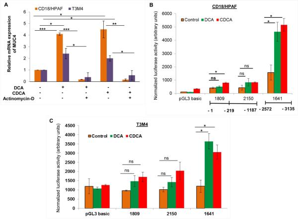
Figure 3. BA-mediated transcriptional regulation of MUC4.
A. After 8h of serum starvation, both CD18/HPAF and T3M4 cell lines were treated for 12h with 50 μM of DCA, CDCA or vehicle control (ethanol) in the presence or absence of actinomycin-D (2 μg/ml). Following treatment, cDNA was prepared from isolated RNA and used for real-time PCR to analyze the quantitative expression of MUC4 gene. The represented graph is demonstrating that inhibition of transcription attenuates DCA- and CDCA-mediated increase in MUC4 expression in both CD18/HPAF and T3M4 cell lines. B. Luciferase assay was performed in CD18/HPAF cell line transfected with MUC4 promoter-truncated constructs, followed by 4h treatment of 50 μM of DCA and CDCA. A significantly elevated luminescence was detected upon BA treatment, primarily at the distal promoter region. C. Similar to CD18/HPAF cells, T3M4 cells also showed significantly elevated luminescence at the distal promoter region upon BA (50 μM) treatment. (*p<0.05, ** p<0.01, *** p<0.001, ns means non-significant)

