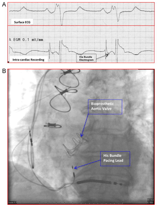
Figure 1.
Intracardiac recording and fluoroscopic appearance of parahisian pacing site. A: Intracardiac electrograms (lower tracing) obtained at a site in the high septum where the His bundle pacing lead was deployed. The His bundle electrogram is indicated by the arrow. The surface electrocardiogram (ECG; upper tracing) is also shown. B: Fluoroscopic appearance of the final position of the permanent His bundle pacing lead and the atrial and ventricular leads in a shallow right anterior oblique projection.

