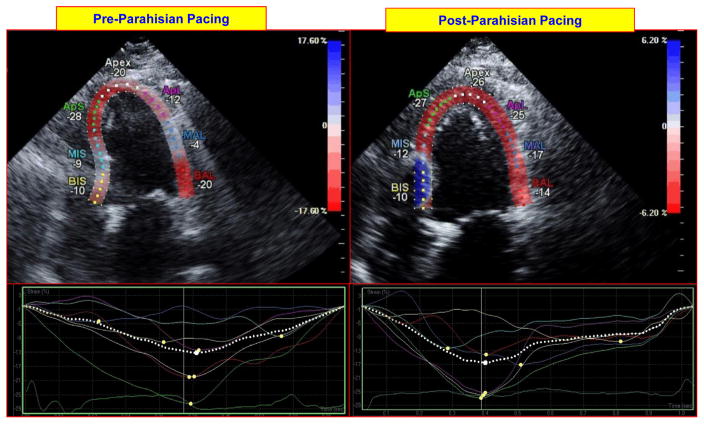
Figure 3.
Impact of parahisian pacing on regional left ventricular (LV) deformation on echocardiographic strain imaging. The top panels on the left and right show longitudinal (3-chamber) images of the left ventricle at peak contraction before and after initiation of parahisian pacing. Regional LV deformation is shown for each region (expressed in negative % deformation). The bottom panels show regional strain profiles of each region before and after parahisian pacing; these are more uniform following pacing. BIS = basal inferoseptal; MIS = mid inferoseptal; ApS = apicoseptum, ApL = apicolateral; MAL = mid anterolateral; BAL = basal anterolateral.

