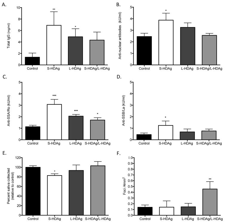
Figure 4.
Expression of HDV antigens recapitulates a Sjögren’s syndrome-like phenotype in an in vivo murine model. (A) Total IgG was significantly increased in mice expressing S-HDAg and L-HDAg, but not in mice that received combination S-HDAg/L-HDAg (t-test, **P<.01, *P<.05, n=7–15). (B) Antinuclear antibodies (ANA) were elevated in mice expressing S-HDAg (t-test, *P<.05, N=6–11). (C) Anti-SSA/Ro60 antibodies were significantly elevated in mice expressing S-HDAg, L-HDAg, and the combination of S-HDAg/L-HDAg (t-test, ***P<.0005, *P<.05, n=7–11). (D) An increase in Anti-SSB/La antibodies was observed in mice that expressed S-HDAg (t-test, *P<.05, n=6–11). (E) Mice were cannulated with adeno-associated virus (AAV) containing expression cassettes for luciferase (control), S-HDAg, L-HDAg, or a combination of the two HDV antigens (S-HDAg/L-HDAg). Mice that expressed S-HDAg showed a significant decrease in pilocarpine stimulated saliva flow compared to controls (t-test, *P<.05, n=6–14). (F) A significant increase in foci was noted in mice that expressed a combination of S-HDAg and L-HDAg (t-test, **P<.005, n=6–14).

