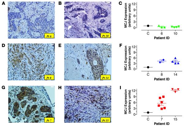
Figure 2. Variable levels of MCJ expression in primary breast cancer biopsies.
(A) Core needle biopsies from breast cancer patients were taken at the time of diagnosis. Sections 3–5 μm in thickness were stained with anti-MCJ mAb and counterstained with hematoxylin. Pictures depict variable intensity of staining among different patient samples; low (patients [Pts.] #6, #10; A and B), intermediate (Pts. #8, #14; D and E), or high (Pts. #7, #15; G and H). Original magnification ×400. (C, F, and I) Relative expression levels of MCJ mRNA isolated from core needle biopsies obtained at diagnosis were compared using qPCR. Each patient sample was run at least in duplicate, and the data shown represent the individual determinations for the indicated patients, as well as the mean ± SEM of the obtained values. Variable levels of MCJ mRNA are depicted from representative patients.

