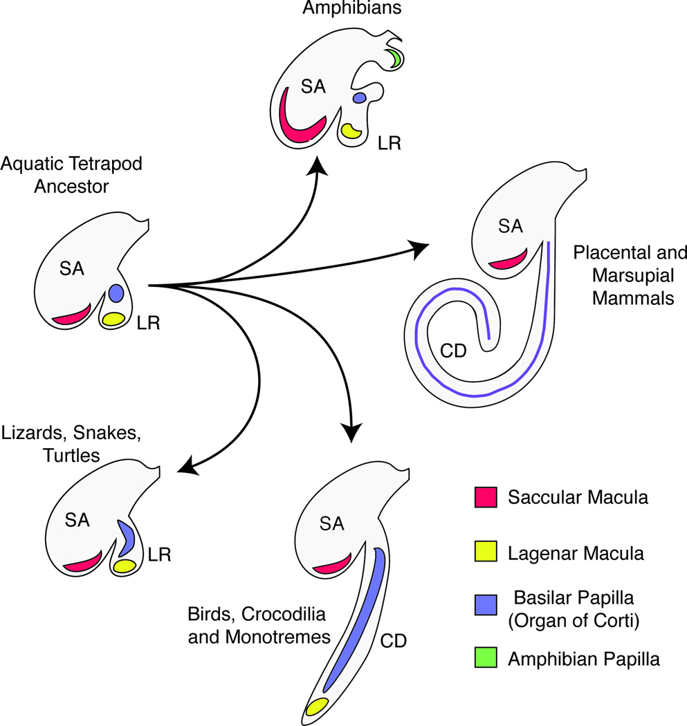
Figure 1.
Evolutionary divergence of the inner ear showing the emergence of the cochlea. The aquatic ancestor of modern tetrapods likely had an evagination of the saccule (SA), termed the lagenar recess (LR) that contained the macula lagena (yellow) and a small basilar papilla (purple). This arrangement is seen today in the coelacanth Latimeria (Fritzsch, 1987, Fritzsch, 2003) and persists to varying degrees in modern lizards, snakes and turtles, and in many modern amphibians which also have a second unique auditory organ, the amphibian papilla (green). In birds, crocodilians and monotremes, the basilar papilla has elongated to different extents, with the lagenar macula being displaced to the distal tip of the cochlear duct. In therian mammals, the lagena has been lost and the elongated basilar papilla (purple) running the length of the cochlear duct is termed the organ of Corti. In each case, only the pars inferior of the inner ear (saccule, lagenar recess and cochlea duct) are shown in the diagram. This diagram is intended to show the basic trends occurring during the evolution of the cochlea, although in reality considerable variation occurs in the shape and size of the sensory organs in each of the main groups shown in the diagram (Fritzsch et al., 2013, Gleich et al., 2004, Manley, 2004, Manley, 2012, Smotherman and Narins, 2004, Vater et al., 2004).

