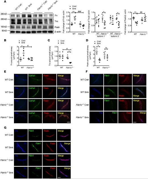
Figure 5. Fbln1c binding proteins are altered in whole lungs and around small airways in WT and Fbln1c−/− mice with experimental COPD.
WT and Fbln1c−/− mice were exposed to cigarette smoke for 8 weeks to induce experimental COPD; controls were exposed to normal air. (A) Fibronectin (Fn), tenascin-c (Tnc), and periostin (Postn) protein levels in whole lungs assessed by immunoblot (left), and fold change of densitometry normalized to β-actin (right). (B) Fn, (C) Tnc, and (D) Postn area around the small airways analyzed by IHC and normalized to perimeter of basement membrane (Pbm). n = 24–40 airways from n = 5–6 mice per group. (E) Colocalization of Tnc (blue), Col1a1 (green), and Postn (red) around the small airways. (F) Colocalization of Tnc (blue), Fbln1 (green), and Postn (red). (G) Colocalization of Fn (blue), Fbln1 (green), and Postn (red) (scale bar: 15 μm. n = 3.). Results are mean ± SEM. *P < 0.05, **P < 0.01 compared with air–exposed WT or Fbln1c−/− controls; #P < 0.05, ##P < 0.01, ###P < 0.001 compared with CS-exposed WT controls. Statistical differences were determined with 1-way ANOVA followed by Bonferroni post-test.

