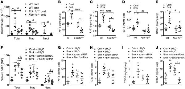
Figure 6. Absence of Fbln1c protects against inflammation in experimental COPD.
WT and Fbln1c−/− mice were exposed to cigarette smoke (CS) for 8 weeks to induce experimental COPD; controls were exposed to normal air. (A) Differential inflammatory cell counts in bronchoalveolar lavage fluid (BALF). (B) TNF-α, (C) IL-33, (D) TGF-β, and (E) CXCL1 protein in whole lungs measured by ELISA. WT mice were treated with Fbln1c or scrambled siRNA from weeks 6–8 of 8 weeks of CS exposure. (F) Differential inflammatory cell counts in BALF. (G) TNF-α, (H) IL-33, (I) TGF-β, and (J) CXCL1 protein in whole lungs. Results are mean ± SEM. *P < 0.05, **P < 0.01, ***P < 0.001, ****P < 0.0001 compared with normal air–exposed WT or Fbln1c−/− controls; #P < 0.05, ##P < 0.01, ####P < 0.0001 compared with CS-exposed WT controls; $P < 0.05 compared with CS-exposed controls treated with scrambled siRNA. Statistical differences were determined with 1-way ANOVA followed by Bonferroni post-test.

