
Figure 1.
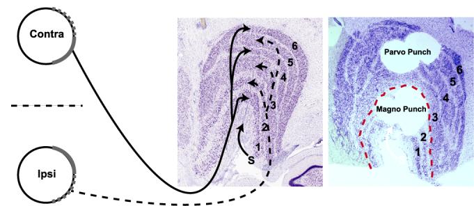
A schematic diagram identifying the target dLGN layers harvested for microarray analysis (Left). Contralateral and ipsilateral retinal ganglion cell axons originate from nasal and temporal retinal ganglion cells respectively and terminate in layers 1, 4 and 6 (contralateral) or 2, 3 and 5 (ipsilateral) of the dLGN, visualized here by Nissl staining. Axons terminating in outer (1 and 2) and inner (3-6) layers form the magno- and parvocellular visual streams respectively. The S layers are indicated (arrow). Magnocellular and parvocellular layers after microdissection with tissue punches is shown by post—hoc Nissl staining (Right).