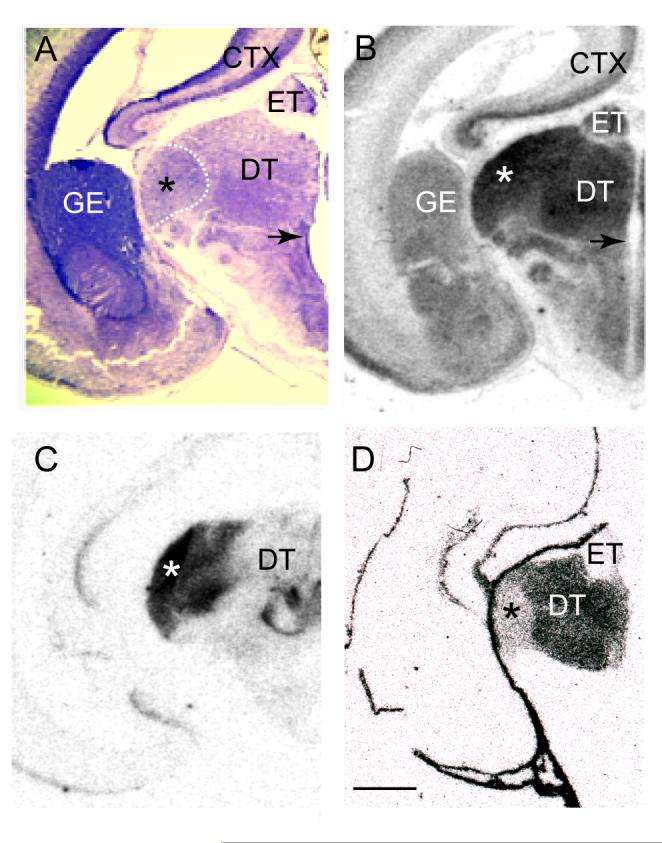
Figure 10.

Markers of dLGN laminae in adult brain are expressed in embryonic day 55 (E55) thalamus. (A) Coronal Nissl stained section through an E55 monkey thalamus illustrating cytoarchitectural differentiation of thalamic nuclei. The location of the dLGN (*) in relation to the anlagen of dorsal thalamus (DT) and epithalamus (ET) is indicated by the dashed bounding box. M, P or K laminae cannot be reliably identified at this age (Huberman et al., 2005). An arrow indicates the location of the third ventricle. (B-D) Autoradiographic images of coronal sections adjacent to that in (A) processed for in situ hybridization with riboprobes specific to the genes NEFL (B), INα (C) and TCF7L2 (D). NEFL and INα are both heavily expressed in the developing dLGN while TCF7L2 displays relatively low levels of expression. Scale bar in (D) = 1 mm (applies to A-D). Abbreviations: CTX, neocortex; DT, dorsal thalamus; ET, epithalamus; GE, ganglionic eminence.
