Figure 5.
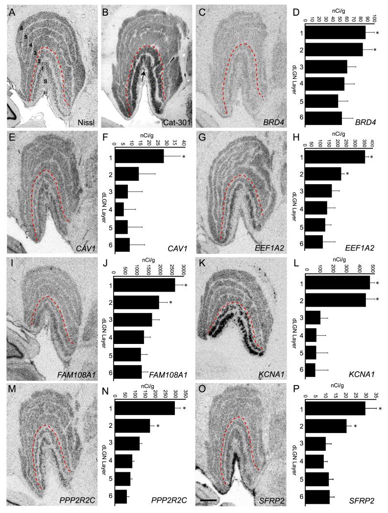
In situ hybridization confirmation of magnocellular enriched gene expression. (A) Coronal Nissl stained section through monkey dLGN illustrating the cytoarchitecture, including the 6 principal cell layers and the S layers (S). The red dashed line indicates the separation between magnocellular (1 and 2) and parvocellular (3-6) layers. (B) Coronal section through monkey dLGN immunostained with monoclonal antibody Cat-301 which recognizes an epitope on the protein aggrecan and is known to be elevated in magnocellular dLGN layers (Hendry et al., 1984). Some staining in S layers is also present (arrow). (C, E, G, I, K, M, O) Images of film autoradiograms illustrating hybridization of radioactive cRNA probes specific to the genes BRD4 (C), CAV1 (E), EEF1A2 (G), FAM108A1 (I), KCNA1 (K), PPP2R2C (M), and SFRP2 (O) on coronal sections of monkey dLGN. Consistent with RT-PCR results each gene displayed elevated expression in one or both magnocellular layers. Laminar differences in mRNA expression were quantified from film autoradiograms of sections processed for in situ hybridization for BRD4 (D), CAV1 (F), EEF1A2 (H), FAM108A1 (J), KCNA1 (L), PPP2R2C (N), and SFRP2 (P). Calibrated densitometric measures were obtained from the principal layers (1-6) of the dLGN. Measures are means ±S.E.M. *, p < .01 (n=5) ANOVA, post-hoc Student’s t-test. Scale bar in O = 1 mm (applies to C, E, G, I, K and M).

