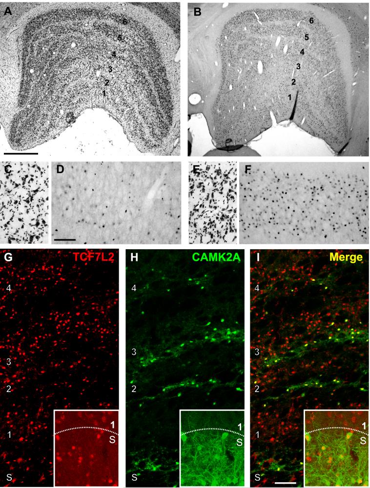
Figure 7.
TCF7L2 protein is enriched in parvocellular, S and intercalated layers of the monkey dLGN. (A) Nissl stained coronal section of monkey dLGN illustrating the principal relay layers (1-6) and the intercalated layers that separate them. The S layers are not readily visible in this section (B) Immunohistochemical labeling of TCF7L2 protein in a coronal section of monkey dLGN adjacent to that in (A). (C) and (E) Higher power images of Nissl stained cells in layer 1 (C) and 6 (E) of dLGN. (D and F) Higher power images from sections adjacent to those in C and E show immunoreactivity for TCF7L2 protein in layers 1 (D) and 6 (F) of dLGN. More immunoreactive cells are observed in layer 6 compared with layer 1. (G-I) Double immunofluorescent labeling of TCF7L2 (red) and type II alpha calcium/calmodulin-dependent protein kinase (CAMK2A) (green) in dLGN. Merged images (I) show colocalization of TCF7L2 and CAMK2A in the intercalated layers of the dLGN and in the S layers (insets). Scale bars = 1 mm in A (applies to A, and B), 250 μM in D (applies to C-F) and 200 μM in I (applies to G-I).

