Figure 9.
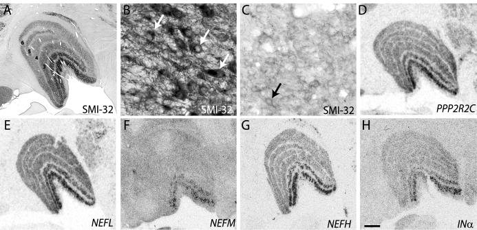
Phosphatase activity and neurofilament expression is greater in magnocellular layers of monkey dLGN. (A) Immunohistochemical staining of a coronal dLGN section with SMI-32 monoclonal antibody showing increased staining of the magnocellular layers (1, 2) of the dLGN compared with parvocellular layers (3-6). (B, C) Higher power images of SMI-32 immunostaining shows denser reaction product over layer 1 (B) compared with layer 6 (C) and illustrate positive immunoreactivity within cell bodies (arrows). (D-H) Autoradiographic images of coronal dLGN sections processed for in situ hybridization showing increased hybridization of riboprobes specific to PPP2R2C (D), NEFL (E), NEFM (F), NEFH (G) and INα (H) mRNA in magnocellular layers of the dLGN. Scale bar = 1 mm in H (applies to A-H).

