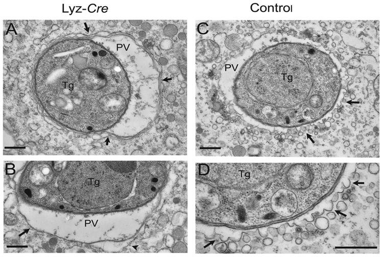
Fig. 4. Electron microscopic analysis of the clearance of T. gondii by IFNγ-activated macrophages.
Macrophages treated as indicated were infected for 5 hours and then analyzed by EM. All images shown are from IFNγ/LPS activated macrophages.
(A) Parasites (Tg) within Atg5 deficient cells, reside within conventional parasitophorous vacuoles (PV) bounded by host ER (arrow heads).
(B) The parasitophorous vacuole membrane is a single unit membrane (arrow) surrounded in places by host ER (arrow head).
(C) Parasites within activated control macrophages are found within vacuoles that are undergoing membrane blebbing and vesiculation (arrows).
(D) Enlarged view from (C) shows membrane blebs protruding from the parasitophorous vacuole membrane (arrows). Scale bars = 0.5 micrometers.

