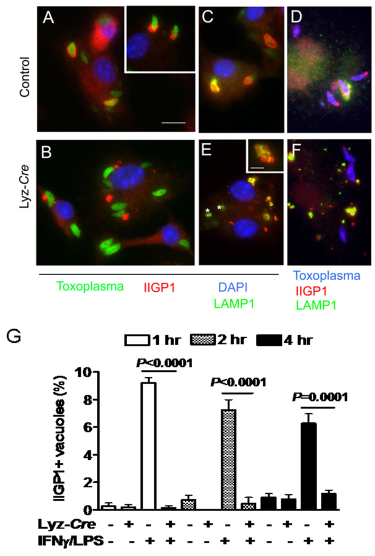
Fig. 6. Atg5 is required for IFNγ/LPS-induced targeting of IIGP1 to the T. gondii parasitophorous vacuole.
Macrophages were stimulated with IFNγ/LPS and infected with T. gondii as in Fig. 2, and then stained for the indicated markers 2 hour after infection. Scale bar = 10 microns for A–F.
(A, B) Localization of IIGP1 (red) with GFP-expressing T. gondii (green) after infection of IFNγ/LPS activated control or Atg5-deficient macrophages. DAPI (blue) staining of nuclei. In control cells IIGP1 decorates the parasitophorous vacuole in a cap, while in Atg5-deficient cells IIGP1 (red) was shunted to large intracellular inclusions and was not recruited to the parasite containing vacuole.
(C) Intracellular fate of GFP-expressing parasites in IFNγ/LPS activated control cells. LAMP-1 (green) was recruited selectively to IIGP1 (red) positive vacuoles and often formed a partial cap, associated with material sloughing from the vacuole surface. DAPI (blue) stains nuclei.
(D) Similar to C except wild type parasites were detected with rabbit anti-Toxoplasma and secondary antibodies conjugated to Alexa355 (blue).
(E) Parasites within Atg5 deficient cells did not recruit IIGP1 or become LAMP1 positive. * GFP expressing parasites. However, IIGP1 positive clusters (red in 1B) were strongly associated with LAMP1. Insert shows enlarged view of intracellular cluster. IIGP1 (red) and LAMP1 (green) were closely associated but not strictly colocalized. (F) Similar to E except wild type parasites were detected with rabbit anti-Toxoplasma and secondary antibodies conjugated to Alexa355 (blue).
G) Quantitation of IIGP1 co-localization with T. gondii parasites in IFNγ/LPS activated macrophages 1, 2, and 5 hours post-infection. Data were collected from two independent experiments counting at least 600 vacuoles.

