
Figure 5. Negative stain EM images of hemagglutinin-Fab complexes.
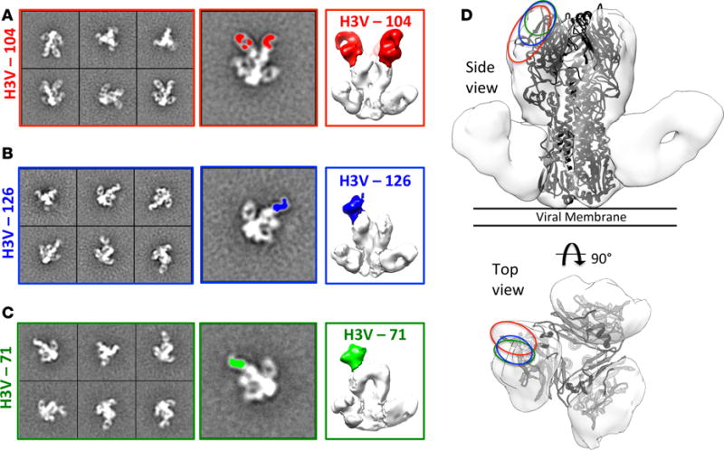
In each case the stem-binding antibody CR9114 was added to the complex in order to improve 3D reconstructions. (A) Reference-free 2D class averages of complex containing Fab 104 (left), single class average with Fab colored in red (middle), and 3D reconstruction (right). (B) Reference-free 2D class averages of complex containing Fab 126 (left), single class average with Fab colored in blue (middle), and 3D reconstruction (right). (C) Reference-free 2D class averages of complex containing Fab 71 (left), single class average with Fab colored in green (middle), and 3D reconstruction (right). (D) Side and top views of HA-Fab 126-CR9114 with Fab 126 removed and crystal structure of H3V (4FNK) fitted. Binding sites of the 3 antibodies described in A–C is highlighted using colors corresponding to Fabs.