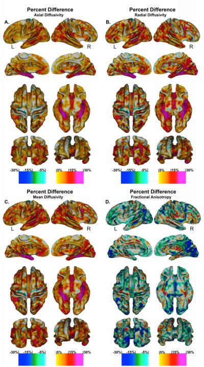
Fig. 5. Percentage Change in Superficial White Matter Diffusivity.
High-resolution vertex based percentage change maps show the change between Controls and Alzheimer’s disease patient’s superficial white matter axial diffusivity at high spatial density. Red colours indicate the percentage diffusivity increased with disease while the blue colours indicated the percentage diffusivity decreased with disease.

