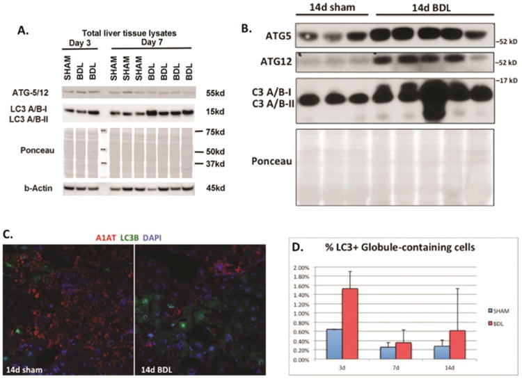
Figure 3. Bile duct ligation induces increased expression of autophagic proteins in PiZZ mouse liver.

A and B. Western blots of total liver tissue lysates showing increased Atg5, Atg12, and LC3A/B after BDL compared to sham livers. Ponceau is shown to confirm equal loading, since cytosketetal changes can affect both actin and tubulin in PiZZ mouse liver. C. Confocal immunofluorescence microscopy showing the majority of LC3B (green) does not co-localize with GC cells labeled with anti-human A1AT (red) antibody (100× magnification). D. Quantitative immunofluorescence data for the LC3 and anti-human A1AT. Graph depicts a small subset of %LC3+ cells co-localizing with ATZ globule-containing cells (normalized to total # of cells).
