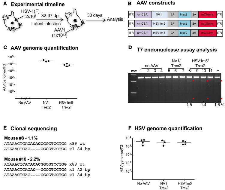
Figure 3. HE-directed mutagenesis of latent HSV in vivo.
(A) Experimental timeline. Mice were infected with 2 × 10⁵ PFU HSV-1(F) in the right eye following corneal scarification and, 32-37 days later, were injected in the right whiskerpad with 1 × 10¹² vector genomes of ssAAV1-smCBA-HSV1m5-Trex2-mCherry or ssAAV1-smCBA-NV1-Trex2-mCherry. Analysis was performed at 30 days after AAV exposure. (B) Schematic representation of the ssAAV constructs used here and Figure 6. (C) Levels of AAV genomes were quantified by ddPCR in right (ipsilateral) TGs from infected mice. Mean ±SD are indicated. (D) Mutagenic event detection by T7E1 assay. The HSV regions containing the target site were PCR amplified from total genomic DNA obtained from the right TG. Products were subjected to T7E1 digestion and separated on a 3% agarose gel. mw, molecular weight size ladder; red asterisks indicate cleavage products. (E) Clonal sequencing of PCR amplicons from TG of treated animals. (F) Levels of latent HSV genomes were quantified by ddPCR in right (ipsilateral) TGs from infected mice. Mean ±SD are indicated.

