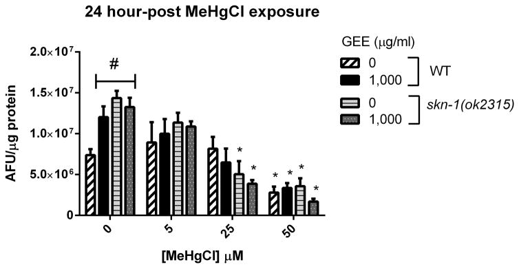
Fig. 5.
ROS-associated fluorescence levels in WT and skn-1 (ok2315) worms untreated and pretreated with GEE 1,000 μg/ml after MeHg exposure and 24-hour recovery. Aliquots of worm’s lysates were incubated for 1 hour with 20 μM DCFDA (final concentration) at 20°C and fluorescence were measured in duplicate in a microplate reader at 485 nm excitation and 520 nm emission wavelengths at room temperature. Data were normalized to protein content. Fluorescence levels were significantly increased in MeHg non-exposed groups in comparison to control and MeHg at 25 and 50 μM significantly decreased fluorescence levels in comparison to MeHg-non-exposed worms of the same group. No significantly differences were detected between untreated and GEE-pretreated worms and between WT and skn-1 (ok2315) worms exposed to MeHg. (#) represents a significantly difference between control and the other groups non-exposed to MeHg by two-way ANOVA followed by Bonferroni’s Multiple Comparison Test (p<0.05) and (*) represents a significantly difference between MeHg-exposed and non-exposed worms of the same group by one-way ANOVA followed by Bonferroni post hoc test (p<0.05). Data are expressed as means of arbitrary fluorescence units (AFU) per μg of protein ± SEM, n=4

