
Figure 2.
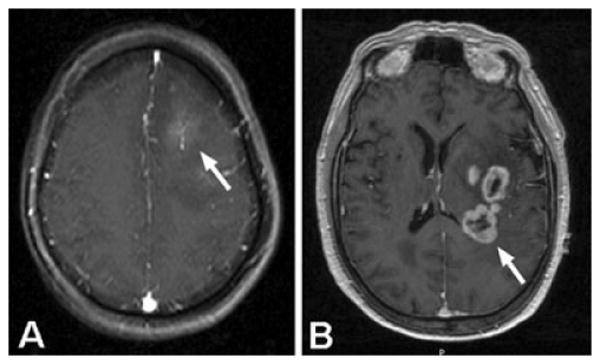
A. Post-contrast, axial MRI of a patient with anaplastic astrocytoma (AA) that showed microscopic evidence of intravascular thrombosis in the biopsy specimen. The MRI shows mild contrast-enhancement of the neoplasm (arrow) with surrounding T1-hypointensity, typical of AA. B. Post-contrast, axial MRI of a patient with glioblastoma, demonstrating the pattern of “ring-enhancement” surrounding central necrosis that is characteristic of this disease (arrow).