
FIGURE 4. Conceptual model of GRK1 docked to Rho*.
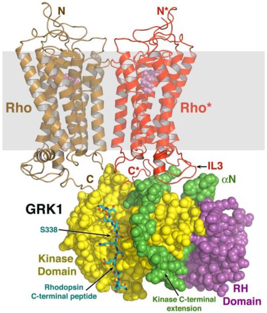
The closed composite model of GRK1 (Fig. 2a) was docked with a model of an array of Rho molecules (Protein Data Bank code 1N3M) (50), of which two molecules are shown here for clarity. GRK1 is rendered as spheres, and the expected lipid bilayer plane is shown as a transparent gray box. A monomer of Rho* (red) was modeled such that its third cytoplasmic loop (IL3) lies close to the proposed receptor-docking site. Using the PKB-GSK3β structure (1O6L) as a guide, the C-terminal peptide of Rho* (carbons are colored cyan, oxygens are red, and nitrogens are blue) was modeled docked to the large lobe, as in Fig. 1d. The GRK1 active site would have easy access to the C-tail of Rho* or of a neighboring unactivated Rho (brown) in the same membrane plane, allowing high gain phosphorylation of ROS.