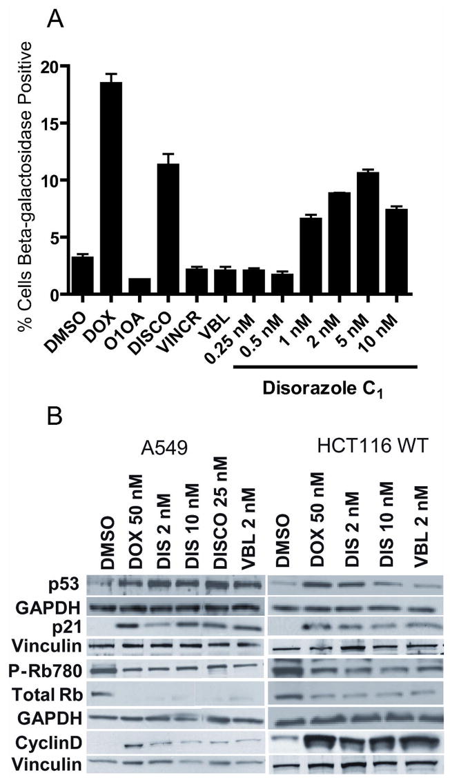
Figure 6. Disorazole C1 causes premature senescence.
(A) A549 cells were treated for 7 days with disorazole C1, IC50 concentrations of discodermolide (DISCO; 25 nM), vincristine (VINCR; 20 nM) or vinblastine (VBL; 2 nM) followed by β-galactosidase staining, which was quantified using the brightfield module and compartmental bioapplication analysis on the ArrayScan VTI with values representative of at least two independent experiments. Doxorubicin (DOX; 50 nM) was used as a positive control for senescence while DMSO vehicle and the inactive analog O1OA (10 nM) were used as negative controls. (B) A549 and HCT116 cells were treated for 7 days and cell lysates were probed by Western blot analysis of protein markers of senescence. GAPDH and vinculin, which were used as loading controls, are located directly below the examined protein lanes.

