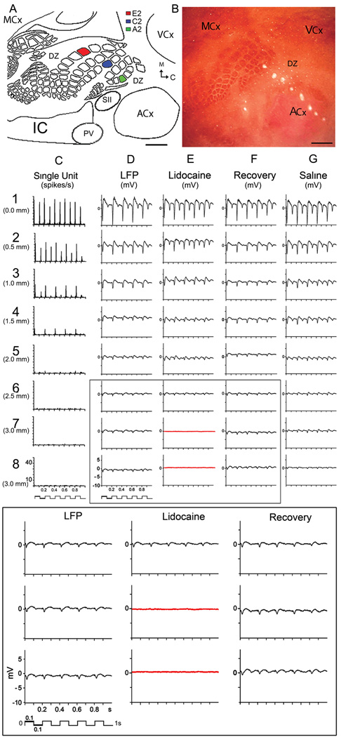
Figure 1. Representative case of evoked SU and LFP responses recorded by the 8-electrode array following whisker A2 stimulation.
A, Schematic based on flattened layer IV CO-stained brain slice illustrating main cortical modalities, their borders, and color-coded barrels of the 3 whiskers studied. B, Lesions produced by the 8-electrode array. Note electrode #1 lesion is localized at A2 barrel and electrodes 4–8 span almost the entire ACx. Scale bars = 1000 µm. C, Evoked SUs decay over cortical distance and completely disappear after electrode 4. D, LFPs also decay over distance but are still present at the last electrode 8. E, LFPs are abolished in electrodes 7–8 (red traces) following lidocaine injection between electrodes 7 and 8, with full recovery after 45 minutes (F). G, Saline injection had no effect. Bottom, Magnification of boxed traces.

