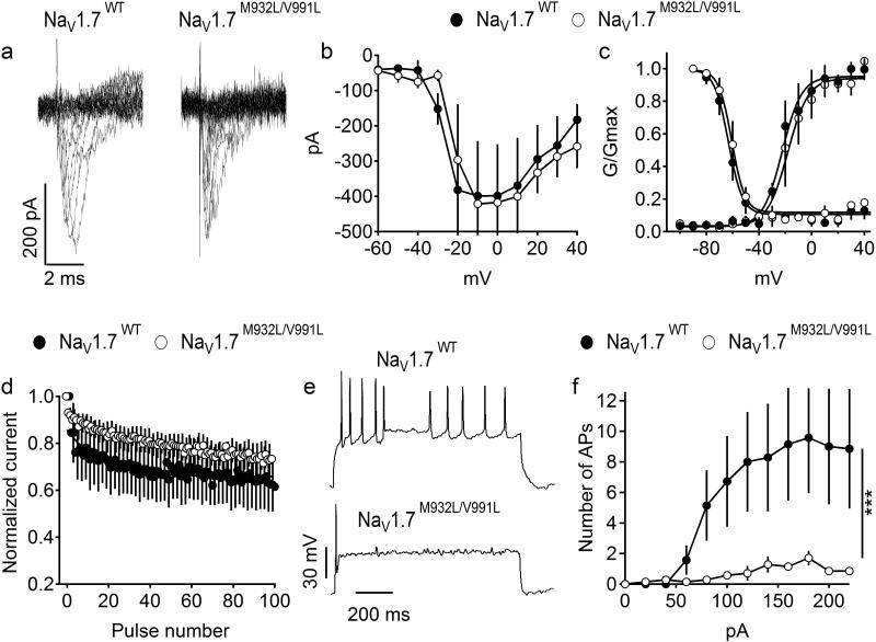
Figure 2. Properties of NaV1.7WT and NaV1.7M932L/V991L expressed in cultured cortical neurons.
(a) Representative set of sodium current traces from cortical neurons expressing NaV1.7WT and NaV1.7M932L/V991L. (b) Mean current-voltage (I-V) relationships of peak currents. (c) Voltage dependence of activation (right curves) and the voltage dependence of steady-state fast inactivation. (d) Mean normalized currents during 100 depolarizations to 0 mV at 50 Hz. NaV1.7WT, n= 4, NaV1.7M932L/V991L, n=5. (e-f) Firing of cortical neurons expressing NaV1.7WT and NaV1.7M932L/V991L NaV1.7M932L/V991L. (e) Representative firing in response to 100 pA depolarizing current injection. (f) Average number of action potentials (AP) in response to 1 s depolarizing current injection at the indicated intensity. NaV1.7WT, n= 7, NaV1.7M932L/V991L, n=7.

