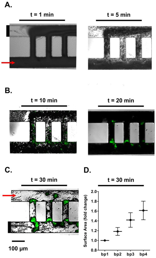
Figure 3. Temporal thrombus growth within ladder network.
DiOC6-labeled whole human blood was perfused at a 2 µL/min flow rate through a PDMS-ladder network coated with collagen and tissue factor; real-time images of thrombus formation were recorded using differential interference contrast, DIC (A) and fluorescence microscopy (B). Networks were subsequently washed with modified Hepes-Tyrodes buffer for 20 min prior to DIC imaging (C). Representative images shown, n = 5. Total surface areas of thrombi per bypass were quantified and normalized to bp1 (D).

