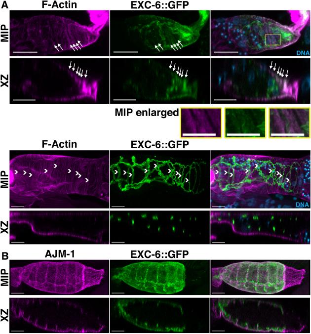
Figure 6. EXC-6::GFP is expressed in the spermatheca and localizes to cell-cell junctions and contractile actin filament bundles.
Shown are maximum intensity projections (MIP) and XZ slices (XZ) reconstructed from confocal Z-stacks. (A) Spermathecae of EXC-6::GFP expressing animals were stained with DAPI to mark DNA and fluorescent phalloidin to mark F-actin. EXC-6::GFP localizes to F-actin bundles near the basal surfaces of the epithelial cells lining the spermatheca (arrows in top spermatheca), and to wavy ribbons closer to the spermatheca lumen (arrowheads in bottom spermatheca). The boxed region of the top spermatheca is enlarged (MIP enlarged) to highlight F-actin bundles with faint associated EXC-6::GFP. (B) Spermathecae of EXC-6::GFP-expressing animals were immunostained for AJM-1. AJM-1 decorates spermatheca apical and lateral junctions, and EXC-6::GFP overlaps with the apical portion of AJM-1. Scale bars, 10 μm.

