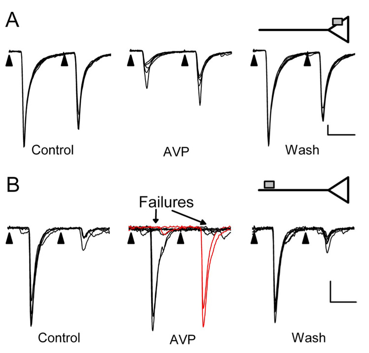
Figure 2.
Two distinct modes of AVP action to inhibit ST transmission. AVP (3 µm) either depressed ST-EPSC amplitudes (A) or induced intermittent ST-EPSC failures (B, arrows). Synaptic transmission rarely failed in control conditions (far left panels, 2 different second-order NTS neurons A and B). The different responses appear mutually exclusive such that increases in ST-EPSC failures had no amplitude changes and vice versa. In failure-type responses, full amplitude EPSC2s were observed when EPSC1 failed during AVP, indicating no previous activation of release sites (B, red current traces). Return to control solution (Wash) reversed AVP actions. These two modes of AVP action are consistent with afferent inhibition either at a presynaptic intraterminal action [A, inset; AVP receptor as gray square directly on an ST terminal (triangle)] or at extraterminal AVP receptors distant from the terminal release site (B, inset). EPSC characteristics: A, 4.3 ms latency, 114µs jitter; B, 7.8 ms latency, 51.5µs jitter. Each trace in A and B displays eight consecutive sweeps overlaid in each condition. Calibration: 10 ms, 100 pA.

