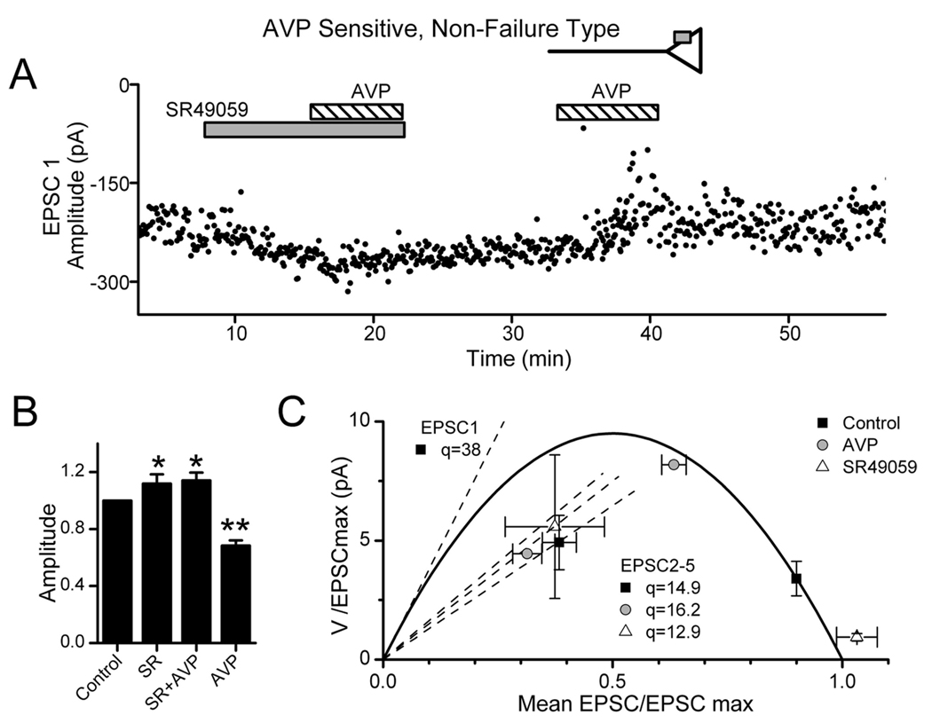
Figure 9.
V1a receptors inhibit ST transmission at second-order NTS neurons. A, During control conditions in this representative neuron (7 ms latency, 132µs jitter), EPSC1 varied over a narrow range of amplitudes. The V1a-selective antagonist SR49059 (gray bar; 10µm) increased EPSC1 amplitude and decreased variance. In the continuing presence of SR49059, added AVP (diagonally striped bar; 3µm) was without effect. Subsequent AVP alone decreased EPSC amplitude and markedly increased EPSC1 variance. B, Summary means for nonfailure, AVP-sensitive neurons tested (n = 4) show that SR49059 (SR) increased the average ST-EPSC amplitude (relative to control) with or without coapplication of AVP compared with control (*p < 0.05 or **p < 0.01,RM ANOVA and paired t test). AVP alone markedly decreased amplitude compared with all conditions. C, In the mean V–M relationship for these neurons (n = 4), SR49059 increased EPSC1M while decreasing V in good agreement with the parabolic V–M model and thus indicates increased release probability when V1a receptors are blocked. AVP alone had the opposite actions and reduced M with increased V as predicted by the parabolic model. These findings indicate a tonic activation of V1a receptors by endogenous AVP. V–M values for EPSC2–5 were linearly related, and the decreased slope of the linear fit indicated decreased quantal size (EPSC3–5 omitted for clarity). In five AVP-resistant second-order NTS neurons tested using identical protocols, EPSC characteristics were unresponsive to AVP or SR49059. None of these treatments altered holding currents ( p > 0.5).

