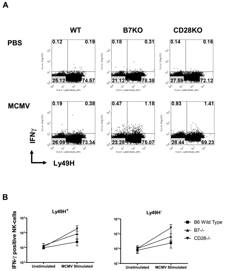
Figure 6. Costimulation and Ly49H+ NK-cell interferon γ (IFN-γ) responses during acute MCMV infection.
Wild type C57BL/6, B7-/- or CD28-/- knockout mice infected with Smith MCMV (5 × 10⁴ pfu) or receiving vehicle (PBS) had splenic tissues removed 2 days after infection. Single splenocytes were prepared, stained with MAb to CD3 (PerCP-Cy5.5), NK1.1 (PE) and Ly49H (APC) followed by intracellular staining with FITC-conjugated MAb to IFN-γ, and then analyzed by four-color flow cytometry. CD3- NK1.1+ cells were gated as described in Figure 3, and further analyzed for Ly49H+ and IFN-γ expression. Ly49H+ NK-cells expressing IFN-γ were enumerated. A. Representative of flow cytometric plots for Ly49H+IFN-γ+ NK-cells from each group. B. Numbers of INF-γ-producing Ly49H+ and Ly49H- NK-cells in each group. Each data point represents mean + standard error for n=3 mice.

