
Fig. 6.
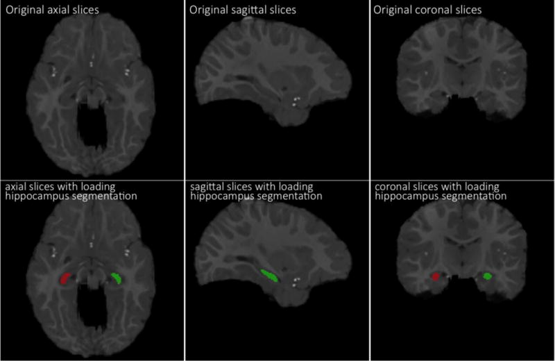
Demonstration of manually-segmented hippocampi in a 12-month-old infant brain MR image. The top row shows the original slices (in axial, sagittal, coronal views), and the bottom row shows manually-segmented left and right hippocampi in red and green.