
Fig. 9.
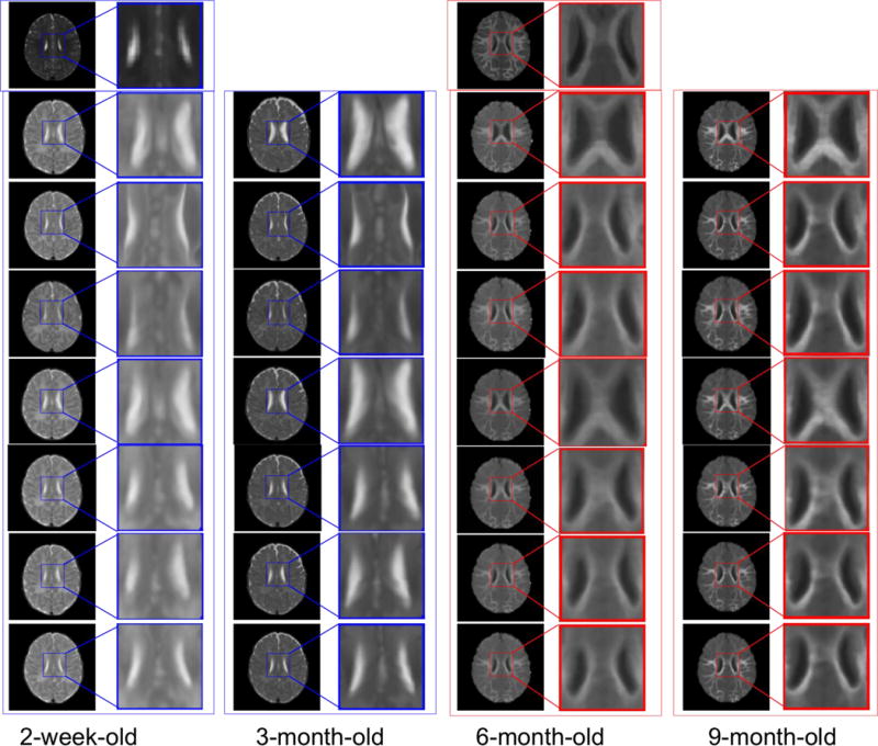
Inter-subject registration results on infant brain images at different time points. The first row shows the 12-month-old template, with T2-weighted image (left) and T1-weighted image (middle). In the whole figure, the blue boxes denote the T2-weighted images, and the red boxes denote the T1-weighted images. From left to right columns, we show the registration results for the 2-week-old, 3-, 6-, and 9-month-old images by linear registration (2nd row), direct use of diffeomorphic Demons (3rd row), CC (4th row), MI (5th row), HAMMER (6th row), diffeomorphic Demons after applying our learned patchwise appearance-displacement model (7th row), and our full method (last row), respectively. Similar to Fig. 7, the 7th row in this figure shows that the anatomical discrepancies are decreased, and the 8th row shows that both anatomical and appearance discrepancies are decreased. Even for inter-subject registration case, as shown in the last row, our proposed method can still obtain good registration results.